
* Paola Rossi
Introduction
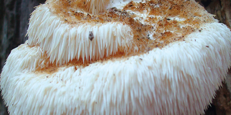
Hericium erinaceus (Bull.:Fr.) Pers. (Hericiaceae, higher Basidiomycetes) is one of the edible and medicinal mushrooms distributed in Asia, Europa and North America. It is one of the wood-destroying fungi that cause white rot; it is a temperate mushroom that requires cool temperatures of 18°C to 24°C to produce fruit bodies (Fig.1).
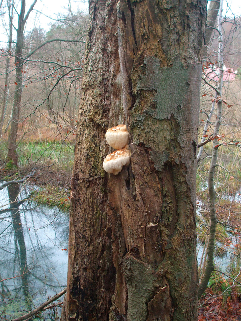
Hericium erinaceus (Hr) is a saprophytic inhabitant on dead trunks of hardwoods, including oak, walnut, beech, maple, sycamore, elm and other broadleaf trees. The nutritional and medicinal properties of Hr are well known in Europe, China and Japan (Chang and Wasser, 2012). In China, it is called Houtou, as its fruit bodies look like the head of a baby monkey, and Shishigashira (Lion’s Head). It is one of the famous four dishes in China (the other three are sea cucumber, bear palm and bird’s nest). In Japan, it is called Yamabushitake because it resembles the ornamental cloth worn by Yamabushi – Buddhist monks practicing asceticism in the mountains.
Hr comprises numerous compounds with intriguing biologic activities; these include promotion of the synthesis of nerve growth factor (NGF) and brain derived neurotrophic factor (BDNF), cytotoxic functions, antimicrobial effects, and antitumor and antioxidant activities. The compounds hericenones C–H and erinacines A–I have been isolated from the Hr fruiting body and mycelium, respectively. Hericenone and erinacine are low-molecular-weight, lipid-soluble compounds that can cross the blood-brain barrier. These compounds have been shown to stimulate NGF synthesis in cultured astrocytes (Kawagishi et al., 1991, 1994, 1996). Moreover, oral administration of Hr increases expression of NGF mRNA in the mouse hippocampus. NGF has been shown to exert crucial functions in the central nervous system (CNS). Results of preclinical studies involving pathological animal models demonstrate that NGF acts on cholinergic neurons, induces neuronal differentiation, promotes neuronal survival and regeneration, and ameliorates neurodegeneration and cognitive deficits.
In mice, oral administration of Hr prevents amyloid β (25-35) peptide–induced impairment of spatial short-term memory and visual recognition memory (Mori et al. 2011). Recently, orally delivered Hr was found to confer neuroprotection in mice that had undergone middle cerebral artery occlusion (Hazekawa et al. 2010).
Despite an extensive investigation of the effects of Hr in cognitive pathological conditions, before us, no one have investigated the effects of dietary supplementation with H. erinaceus in healthy mice. Thanks to the scientific collaboration between Miconet, an academic spin-off of the University of Pavia and AVD Reform s.r.l. (Casal Noceto, Pavia) we assessed the study of the effect of the dietary supplement “Micotherapy Hericium” on central nervous system. The AVD Reform supplement “Micotherapy Hericium” contains mycelium and fruiting body extract of Hr in a ratio 4/1.
We investigated the effect on behavior and on the neuronal network involved in memory skills in wild-type healthy mice. A group of wild-type mice were fed for two months with a dextrin dietary supplementation and another one with Hrdietary supplementation. We performed in vivo and in vitro experiments to assess novelty exploration and recognition memory after Hr dietary supplementation (Brandalise et al. 2017; Rossi et al. 2018). We also presented some preliminary data on Hr supplementation on mood and sleep disorders in a group of overweight female subjects treated with hypocaloric diet.
Where is located memory in our brain?
Memory is located in a part of our brain called medial temporal lobe comprising hippocampus and parahippocampus regions. The hippocampus belongs to the limbic system and plays important roles in the consolidation of information from short-term memory to long-term memory and spatial navigation. Humans and other mammals have two hippocampi, one in each side of the brain. In Alzheimer’s disease, the hippocampus is one of the central nervous system region to suffer damage; memory loss and disorientation are included among the early symptoms. Damage to the hippocampus can also result from hypoxia, encephalitis, or medial temporal lobe epilepsy. People with extensive, bilateral hippocampal damage may experience anterograde amnesia – the inability to form or retain new memories.
Memory to a cellular level is expressed in the neuronal network as neural plasticity. Long term plasticity known as long-term potentiation (LTP) is the cellular mechanism at the basis of memory storage in our brain.
Recognition memory
Our memory can be distinguished in two major types (Fig.2).
– Implicit memory (or non-declarative), is a memory that relates to the mode of “Executing an act” and it is invoked to the mind unconsciously. This kind of memory is generally connected with the performance-based training, motors or perceptions, of reflex type. It is a rigid memory (not ductile), closely related to the original conditions in which learning tooks place;
– Explicit memory (or declarative) is a memory that involves knowledge of facts about people, places, or objects. This type of memory is evoked in mind with deliberate and conscious efforts. It is a very flexible memory and requires the ability to associate together different informations.
The expression of declarative memory depends on connected anatomical structures located in the medial temporal lobe of the central nervous system, in particular hippocampus and parahippocampal cortex. Declarative (or explicit) memory is further subdivided into episodic and semantic memory. Episodic memory is defined as a memory of events personally experienced. People remembers his past with the consciousness of “where” and “when” events have taken place (Fig. 2).
Knowledge of facts and notions, independently by personal experiences, is defined semantic memory. An example is the notions we learned from school or books. The semantic memory includes the knowledge of things, facts and concepts, as well as words and their meaning, independently by the spatio-temporal context.
The recognition memory is a type of episodic memory and concerns autobiographical experiences and often includes the spatio-temporal context. One of the features highlighted in people with cognitive decline, such as patients suffering from Alzheimer’s, is the difficulty to recognize a familiar object, previously encountered and the relative difficulty in distinguishing it from a new object. The memory of “recognition” is a fundamental aspect of our ability to remember and to identify an event. Researchers have long been interested in the mechanisms underlying recognition memory and currently there is a general agreement that two distinct cognitive operations, recollection and familiarity, contribute to recognition memory. The recognition process is therefore composed by at least two components (Brown et al., 2001, Fig 3):
– Discrimination of the familiarity, that is, knowledge of what has been previously encountered;
– Ability to remember information about the spatial and /or temporal context in which objects were encountered.
We can make an easy example to better understand this point. By meeting a person on the road, we can recall the name (“Familiarity”) and we can recall where we have met him before (“remember informations”). The two processes are therefore the remember as a familiar people and the recall of the previous context encountered.
This two components of recognition memory could be localized in the same neuronal network or could be localized in two different regions of the central nervous system (Brown M.W. et al., 2001)
Ongoing debate which is central to our understanding of recognition memory function is whether recollection and familiarity reflect different memory processes that can be dissociated anatomically and functionally (dual-processes model) or it is a single unitary process expression of memory traces of different strength in the context of a unitary declarative memory system (unitary-strength models, Ameen-Ali et al., 2015). In the first hypothesis, recognition memory could be supported by two functionally distinct processes mediated by different structures in the medial temporal lobe; the parahippocampal region, could be part of a circuit involved in familiarity and recognition of individual items and the hippocampus, supporting recollected associations and relationships amongst stimuli (for a review, see Ameen-Ali et al., 2015). Studies involving human amnesic patients with hippocampal damage have provided useful insight into this debate and it has offered support to the dual-process model. In animals, lesion studies using the spontaneous novel object recognition task (NOR) and object-location task (O-L) have provided a useful insight into the anatomical basis for recognition memory. The perirhinal cortex is critical for successful performance on NOR, while lesions of hippocampal or fornix have no detrimental effect on NOR test. Conversely, rats with dorsal hippocampal lesions cannot successfully perform the OL, while perirhinal cortex lesions have no effect on OL task performance.
Effect of Hericium erinaceus in memory cognitive performances in mice
To explore the role of Hr in vivo in recognition memory and in exploration behavior we evaluated the effect of two months oral supplementation with “Mycotherapy hericium” in healthy mice by using two batteries of spontaneous behavioral tests, the NOR and the Emergence test, from one side and the OL and Ymaze from the other side. All the experiments were recorded with a video camera.
As described before, the recognition memory, a form of declarative memory, can be defined as the ability to discriminate the novelty or familiarity of previous experiences by identifying when something (e.g., an object or an environment) has already been encountered.
The Novel Object Recognition test (NOR, Fig. 4) has become a widely used model for working memory, attention and preference for novelty in rodents. The NOR task evaluates the rodents’ ability to recognize a novel object in the environment. Basically, in the NOR task, there are no positive or negative reinforcers, and this methodology assesses the natural preference for novel objects displayed by rodents. The task procedure consists of three phases: habituation, familiarization, and test phase. In the habituation phase, for the first two days each animal is allowed freely exploring the open-field arena in the absence of objects for 10 minutes. The animal is then removed from the arena and placed in its holding cage. On the third day, during the familiarization phase a single animal is placed in the open-field arena containing two identical sample objects, for 5 minutes. After a retention interval (15 minutes), during the test phase, the animal is returned to the open-field arena with two objects, a familiar one and a new one.
The Emergence test is a variant of the open field test, commonly used for qualitative and quantitative measure of general locomotor activity and of willingness to explore in rodents. The apparatus consist in a typical cage for rodents where we have made a hole through which the mouse was able to get out and explore the environment.
Our data demonstrate that wild-type mice supplemented with Hr increased their recognition memory and the exploration of a novel environment and a novel object (Brandalise et al., 2017). The results of the NOR task indicate that the Hr mice spent more time approaching the novel object than the familiar object; the decreased latency of the first approach and the increase in the frequency of approaches, combined with the longer duration of approaches, further support a state of increased novelty exploration behavior. Conversely, the NOR test revealed no differences between Hr and dx mice in the exploration of the familiar object, indicating that the increase in exploratory activity is specifically oriented to the novel object.
The ability to cope with novelty is essential in all mammal species. Novelty-seeking has been identified as one of the six major human personality dimensions, whereas neophobia describes hesitancy to engage with novel objects and places and can be considered a risk factor for anxiety disorders. A low level of exploratory activity towards novelty is interpreted as a sign of anxiety-like behavior. Furthermore, reduced novelty-seeking and, in turn, increased neophobia can be considered core symptoms of depression; these behaviors are closely related to rigid evaluative patterns and reduced flexibility that also characterize the depressive state. A recently published paper described the reduction of depression and anxiety by 4 weeks’ intake of Hr dietary supplementation in 30 female subjects (Nagano M. et al., 2010).
To explore the role of Hr in spatial memory we evaluated the effect of two months oral administration in wild-type mice by using the Object Location test and Y-maze test.
Object location test (OL, Fig. 5) is a variant of the NOR test and involves multiple items and contextual associations. The task procedure is similar to the NOR test but, after a retention interval (15 minutes), during the test phase, the animal is returned to the open-field arena with two objects, where one is left in the same position while the other is moved to a new position.
In the O-L task, all parameters measured after Hr oral supplementation i.e. number, total duration, average duration of approaches and latency of first approach are not statistically significant suggesting that in wild-type mice Hr has no effect on spatial working memory.
The Y maze is a simple and automatable task that allows for rapid assessment of the willingness of rodents to explore new environments. Rodents typically prefer to investigate a new arm of the maze rather than returning to one that was previously visited. Because the behavior is not reinforced with external rewards or punishments, this task is considered spontaneous. In Y-maze task is possible to measure the locomotor activity in terms of the frequency of arm entries, spontaneous alternation and spatial working memory. Many parts of the brain, including the hippocampus, septum, basal forebrain, and prefrontal cortex are involved in this task.
Data obtained in Hr supplemented mice performing OL test were confirmed in Y-maze test that reveals that the alternation percentage, a measure of the mouse performance in choosing different arm at each inspection, did not change. In supplemented mice an increase in general locomotion activity was revealed by the number of total arm entries. The lack of effects with O-L and Y-maze tasks that has been described to be hippocampal-dependent and, on the other side, the increase in recognition memory obtained with NOR and emergence tasks that has been described parahippocampal-dependent, strongly suggests that Hr supplementation has a selective role in increasing parahippocampal cognitive performances. An other outcome coming from our studies is that the effects of Hr supplementation in wild-type animals strongly suggest that the recognition memory is a dual-process implying different behavioral mechanism and/or different anatomical structures.
Hericium erinaceus effect on hippocampal network
We studied the effect of Hr in supplemented wild-type mice to a network and cellular level on hippocampus brain slices by performing electrophysiology recording in the mossy fiber-CA3 region, a glutamatergic synapse of the hippocampus.
We recorded spontaneous and evoked excitatory post-synaptic currents in mice treated with two months of oral administration with dextrin (as placebo control) or Hr. Recordings were obtained by using whole-cell voltage-clamp technique in hippocampus CA3 pyramidal neurons.
We demonstrated that after 2 months of oral supplementation with Hr there a higher amount of neurotransmitter is released from mossy fiber to CA3 pyramidal neurons. This conclusion was supported by higher frequency and higher amplitude of spontaneous excitatory activities recorded in pyramidal neurons and lower number of stimulation failures and decreased pair-pulse ratio of evoked activities in Hr-fed mice (Brandalise et al. 2017, Rossi et al. 2018). Therefore, after oral supplementation, Hr increase glutamatergic neurotransmission in the hippocampus structure demonstrating, for the first time, the increase in neurotransmitter release and of neuronal excitatory activity on circuit involved in consolidating memory traces.
Hericium erinaceus effects on human
Hr increases nerve growth factor (NGF) expression in central nervous system, and, in particular in the hippocampus structure. It is assumed that functional deficiency of NGF is related to Alzheimer’s disease and plays a part in the etiology of the disease process. It is known that NGF levels are decreased in the basal forebrains of Alzheimer’s disease patients, and in the frontal cortices of patients with senile plaques (Mori et al., 2008).
Two recent papers describe the oral supplementation in human. In the first, the efficacy of oral administration of Hr was reported in patients diagnosed with mild cognitive impairment, using a cognitive function scale based on the revised Hasegawa Dementia Scale. Laboratory tests were performed to confirm the safety of the supplementation and the absence of adverse effect. The study of Mori et al. (Mori et al. 2009) suggest that continuous intake of foods that promote NGF synthesis may be one of the effective ways to prevent or alleviate Alzheimer’s disease. Consequently, Hr can be regarded as a useful food for the prevention of dementia without any adverse effects.
A second study on human has described an effective action of Hr in improving mood disorders in a group of women suffering from depressive and anxious symptoms (Nagano et al. 2010).
We assessed the effects of Hr supplementation on mood and sleep disorders in a group of overweight female subjects treated with hypocaloric diet.
We recruited subjects who had both mood disorders and a condition of overweight-obesity (BMI> 25) at the Policlinico Ca’ Grande at the Obesity Center of Milan. Mood disorders were discriminated on the scores analysis in the self-evaluation tests for anxiety, depression, emotional eating and sleep disorders, routinely administered to all patients at the obesity center. Hr was taken for 8 weeks by the subjects included in the treatment group and the neurotrophic activity was evaluated through the scores of the self-evaluation tests compared to the control group under the same low-calorie diet. The preliminary data obtained suggest that Hr supplement “Mycotherapy Hericium” could be used for the prevention and treatment of obesity and depression.
Conclusion
Our data indicate that Hr supplementation with in healthy mice increase the recognition memory of new object and of new place, an antineophobia behavior. No effect were seen on spatial memory tasks, demonstrating that Hr oral supplementation is specifically addressed to parahippocampal region increasing glutamatergic synaptic drive, novelty exploration behavior, and recognition memory.
This result is paralleled by an increase in spontaneous and evoked glutamatergic neurotransmission at the mossy fiber-CA3 synapse in hippocampus. In human Hr supplementation with “Mycotherapy hericium” decreases mood and sleep disorders in overweight women under a low caloric diet.
This data on the effects on brain of Hericium erinaceus oral supplementation both in mice and in human are really promising and yielded several key findings that we hope will pave the way for new studies in healthy humans in order to bridge the gap between the millenary Eastern medicine and our Western medicine (Wasser S.P., 2014).
* UNIVERSITY OF PAVIA
Department of Biology and Biotechnology “L. Spallanzani”
Miconet, Academic spin-off of the University of Pavia, Italy
References
Ameen Ali K.E. Easton A. Eacott MJ (2015) Moving beyond standard procedures to asses spontaneous recognition memory 37-51 in Neuroscience and Biobehavial Reviews 2015, 53.
Brandalise F, Cesaroni V, Gregori A, Repetti M, Romano C, Orrù G, Botta L, Girometta C, Guglielminetti ML, Savino E, and Rossi P. Dietary Supplementation of Hr Increases Mossy Fiber-CA3 Hippocampal Neurotransmission and Recognition Memory in Wild-Type Mice. Evidence-Based Complementary and Alternative Medicine, Volume 2017 (2017),1-13.
Chang ST, Wasser SP. The role of culinary-medicinal mushrooms on human welfare with a pyramid model for human health. Int J Med Mushrooms, 2012;14(2):95-134.
Ennaceur A. Neave N. Aggleton J.P. (1997) Spontaneous object recognition and location memory in rats: the effects of lesions in the cingulate cortices, the medial prefrontal cortex, the cingulum bundle and the fornix 509-519 in Exp Brain Res 1997
Kawagishi H., Ando M., Sakamoto H., Yoshida S., Ojima F., Ishiguro Y., Ukai N., Furukawa S. Hericenones C, D and E, stimulators of nerve growth factor (NGF)-synthesis, from the mushroom Hericium erinaceus. Tetrahedron Letters; 1991;4561–4564.
Kawagishi H., Shimada A., Shirai R., Okamoto K., Ojima F., Sakamoto H., Ishiguro Y., Furukawa S. Erinacines A, B and C, strong stimulators of nerve growth factor (NGF)-synthesis, from the mycelia of Hericium erinaceum. Tetrahedron Letters; 1994;1569–1572.
Kawagishi H., Shimada A., Hosokawa S., Mori H., Sakamoto H., Ishiguro Y., Sakemi S., Bordner J., Kojima N., Furukawa S. Erinacines E, F, and G, stimulators of nerve growth factor (NGF)-synthesis, from the mycelia of Hericium erinaceum. Tetrahedron Letters 1996;7399–7402.
Hazekawa M, Kataoka A, Hayakawa K, et al. “Neuroprotective effect of repeated treatment with Hericium erinaceum in mice subjected to middle cerebral artery occlusion”. Journal of Health Science, vol. 56, no. 3, pp. 296–303, 2010.
Mori K, Inatomi S, Ouchi K, Azumi Y, and Tuchida T. “Improving effects of the mushroom Yamabushitake (Hr) on mild cognitive impairment: a double-blind placebo-controlled clinical trial”. Phytotherapy Research, vol. 23, no. 3, pp. 367–372, 2009.
Mori K, Obara K and Hirota M, et al. “Nerve growth factor inducing activity of Hr in 1321N1 human astrocytoma cells,” Biological and Pharmaceutical Bulletin, vol.31, no. 9, pp. 1727–1732, 2008.
Mori K, Obara Y, Moriya T, Inatomi S, and Nakahata N. “Effects of Hr on amyloid 𝛽(25–35) peptide induced learning and memory deficits in mice”. Biomedical Research, vol. 32, no. 1, pp. 67–72, 2011.
Nagano M, Shimizu K, Kondo R, et al., “Reduction of depression and anxiety by 4 weeks Hr intake”Biomedical Research, vol. 31, no. 4, pp. 231–237, 2010.
Rossi P., Cesaroni V., Brandalise F., Occhinegro A.,Ratto D., Perrucci F., Lanaia V., Girometta C., Orrù G., and Savino E. Dietary supplementation of Hr on spatial memory in wild-type mice. Accepted in International Journal of Medicinal Mushrooms.
Natural 1